comparative anatomy of dog and horse forelimb
Comparative anatomy of forelimb of camel , ox and horse. Box 5-2Types of Joints Ox: Ulna runs the full length of the radius. 2. Forelimb and thoracic limb may be used interchangeably. The carpus normally has greater than 180 degrees of extension. Biologists use the Atlantoaxialarticular surfaces The dog has an anconeal process, which is near the attachment site of the anconeus muscle. The word canine is an adjective and the word dog is a noun; these terms are used in this consistent grammatical form throughout the chapter. The axis has a dens, which projects cranially to allow pivotal motion between the atlas and axis. Hemal arches are separate bones that articulate with the ventral surfaces of the caudal ends of the bodies of Cd4-Cd6. The canine distal radius has distinct facets for articulation with carpal bones, providing stability in weight bearing. Figure 5-7 Skeleton of the left dorsal (A) and left palmar (B) forepaw of the dog. homologies of vertebrate forelimbs. A normal amount of glide occurs in normal functioning joints. Dogs: Ulna and Radius are NOT fused together. Joint Motion and Shape of Articular Surfaces The human stands upright on the feet, with the plantar aspect of the feet contacting the floor and adjacent to each other. The major direction of motion, such as flexion of the stifle, is physiologic or osteokinematic motion. Comparative anatomy between dogs and humans has been described in other sources. The tarsus, or hock, consists of the talus, calcaneus, a central tarsal bone, and tarsal bones I to IV (see Figure 5-10). This type of stance is termed a digitigrade stance. Because dogs are quadruped, there is weight bearing on all four limbs. The spine consists of five areas of the vertebral column: the cervical vertebrae and its articulation with the head, Forearm or antebrachium: Elbow to carpal joint, One sesamoid bone in the tendon of the abductor pollicis longus, Digits or phalanges I to V, numbered medial to lateral, Dewclaw or pollex or digit I with 2 phalanges, Pads on the paws or digital pads: Weight-bearing pads, Ungual process: Extension of the phalanx into the claw, Dewclaw or digit I or halluxmay be absent, fully developed and articulating with a metatarsal, or may be a vestigial, that is, a trace or rudimentary structure, with a terminal phalanx and no proximal phalanx or metatarsal bone, Digital pads or pads on the hindpawsweight-bearing pads, Ungual process: Extension of the distal phalanx into the nail, Bones in the dog skeleton (excludes auditory ossicles), Pelvic girdle: Right and left hip bones and sacrum, Pelvic complex: Hip bones, lumbar spine, sacral spine, caudal spine, sacroiliac joints, and hip joints, Detailed skeletal anatomy of the atlas and axis from a craniolateral view (, Detailed skeletal anatomy of T6 vertebra from a lateral view (, Detailed skeletal anatomy of the sacrum from a caudolateral view (. Hyoid bone: 1 The canine atlas, or C1 vertebra (see Figure 5-12), has a transverse foramen in each transverse process, a craniodorsal arch, and right and left lateral vertebral foramina, Thoracic vertebrae (see Figure 5-13) have small bodies relative to the size of the entire vertebrae. Scapula Humerus Radius and ulna Manus includes Carpus Metacarpus digits. Dogs are digitigrade animals and bear weight on digits II to V, with the main weight bearing occurring on digits III and IV. The canine axis is very large relative to the size of other canine cervical vertebrae. The accessory carpal bone is not as prominent a structure as in the dog.  The tarsus, or hock, consists of the talus, calcaneus, a central tarsal bone, and tarsal bones I to IV (see Figure 5-10). At T10, the size of the body begins to increase and the length of spinous process decreases. An axis of rotation for a joint motion is a straight line or rod that is 90 degrees to the plane of motion. The ribs have vertebral attachments (see Figure 5-11). Dogs have an abbreviated clavicle that does not articulate with the rest of the skeleton. Dogs have much more limitation in motion in the dorsal and transverse planes. For example, cranial movement of the tibia on a stable femur is named stifle joint extension. Costovertebral Synchondrosis: Costochondralribs with cartilage
  The canine hindlimb is known also as the pelvic limb or rear limb, but we use the term hindlimb. The forelimbs bear 60% of The main planes of motion for dogs are as follows (see Figure 5-1): The sagittal plane divides the dog into right and left portions. The dog stands upright on digits or phalanges of each forepaw or manus and each hindpaw or pes (Figure 5-1). The forelimb skeleton consists of the thoracic or pectoral girdle and bones of the forelimb (see Figures 5-5 and 5-6 ). The terminology used in dogs is consistent with naming flexion as described previously. Pad surface on MCP joints in interosseous tendons of digits II to V; two per digit; smaller The anconeal process is needed for stability in weight bearing. Because the term foot can be interpreted as a front foot or a hind foot, this term is clarified when used or specified as forepaw or manus, or hindpaw or pes. At the talocrural joint, two convex ridges of the trochlea of the talus articulate with two reciprocal concave grooves of the cochlea of the tibia. Directional terms include cranial, caudal, rostral, dorsal, palmar, plantar, medial, and lateral. Condyloid: Atlantooccipital
Caudal and cranial articular surfaces are oriented close to the dorsal plane. Axes of Rotation Glides are shear type or sliding motions of opposing articular surfaces. The hindlimbs bear 40% of the dogs weight. Glides are shear type or sliding motions of opposing articular surfaces. Ligamentous and other soft tissue around the joint guide and restrict the motion that would be possible based on articular surface shape alone. The transverse processes are plate-like and flattened dorsoventrally. The anconeal process is needed for stability in weight bearing. Caudal and cranial articular surfaces are oriented between the dorsal and transverse planes to facilitate cranial and caudal glides needed for cervical spine flexion and extension. It articulates distally with the ulnar carpal and accessory carpal bones by two distal facets and does not have an articular disk. Forelimb and thoracic limb may be used interchangeably. Flexion may also be referenced to limb motions involving closing angles during the swing phase of gait. Dogs are digitigrade animals and bear weight on digits II to V, with the main weight bearing occurring on digits III and IV. The spinal cord ends at lumbar (L) L6-L7. Skeleton of the lateral forelimb of the dog. The carpus normally has greater than 180 degrees of extension. Webcomparative anatomy, the comparative study of the body structures of different species of animals in order to understand the adaptive changes they have undergone in the course of evolution from common ancestors. Physiologic motion in joints with opposing concave and convex articular surfaces involves both roll and glide. Anatomic name: pollex for digit I Caudal and cranial articular surfaces are oriented close to the dorsal plane. The cranial articular surfaces are similar to those in more cranial vertebrae in shape and location; however, the caudal articular processes are bifid and are more centrally located, whereas articular processes in more cranial vertebrae are located more laterally. Dorsal and palmar on DIP joints of digits I to V; cartilage; small It is an ossification in the quadriceps femoris muscle. In the spine, flexion occurs as the back or neck arches dorsally (i.e., the convex portion of the arch is directed dorsally). Types of joints are listed in Box 5-2. Compressive or approximation accessory motions are compressive or pushing-together movements between bones. During extension, the limb reaches out, the digit is extended, and the back or neck is less arched dorsally or arched ventrally.
The canine hindlimb is known also as the pelvic limb or rear limb, but we use the term hindlimb. The forelimbs bear 60% of The main planes of motion for dogs are as follows (see Figure 5-1): The sagittal plane divides the dog into right and left portions. The dog stands upright on digits or phalanges of each forepaw or manus and each hindpaw or pes (Figure 5-1). The forelimb skeleton consists of the thoracic or pectoral girdle and bones of the forelimb (see Figures 5-5 and 5-6 ). The terminology used in dogs is consistent with naming flexion as described previously. Pad surface on MCP joints in interosseous tendons of digits II to V; two per digit; smaller The anconeal process is needed for stability in weight bearing. Because the term foot can be interpreted as a front foot or a hind foot, this term is clarified when used or specified as forepaw or manus, or hindpaw or pes. At the talocrural joint, two convex ridges of the trochlea of the talus articulate with two reciprocal concave grooves of the cochlea of the tibia. Directional terms include cranial, caudal, rostral, dorsal, palmar, plantar, medial, and lateral. Condyloid: Atlantooccipital
Caudal and cranial articular surfaces are oriented close to the dorsal plane. Axes of Rotation Glides are shear type or sliding motions of opposing articular surfaces. The hindlimbs bear 40% of the dogs weight. Glides are shear type or sliding motions of opposing articular surfaces. Ligamentous and other soft tissue around the joint guide and restrict the motion that would be possible based on articular surface shape alone. The transverse processes are plate-like and flattened dorsoventrally. The anconeal process is needed for stability in weight bearing. Caudal and cranial articular surfaces are oriented between the dorsal and transverse planes to facilitate cranial and caudal glides needed for cervical spine flexion and extension. It articulates distally with the ulnar carpal and accessory carpal bones by two distal facets and does not have an articular disk. Forelimb and thoracic limb may be used interchangeably. Flexion may also be referenced to limb motions involving closing angles during the swing phase of gait. Dogs are digitigrade animals and bear weight on digits II to V, with the main weight bearing occurring on digits III and IV. The spinal cord ends at lumbar (L) L6-L7. Skeleton of the lateral forelimb of the dog. The carpus normally has greater than 180 degrees of extension. Webcomparative anatomy, the comparative study of the body structures of different species of animals in order to understand the adaptive changes they have undergone in the course of evolution from common ancestors. Physiologic motion in joints with opposing concave and convex articular surfaces involves both roll and glide. Anatomic name: pollex for digit I Caudal and cranial articular surfaces are oriented close to the dorsal plane. The cranial articular surfaces are similar to those in more cranial vertebrae in shape and location; however, the caudal articular processes are bifid and are more centrally located, whereas articular processes in more cranial vertebrae are located more laterally. Dorsal and palmar on DIP joints of digits I to V; cartilage; small It is an ossification in the quadriceps femoris muscle. In the spine, flexion occurs as the back or neck arches dorsally (i.e., the convex portion of the arch is directed dorsally). Types of joints are listed in Box 5-2. Compressive or approximation accessory motions are compressive or pushing-together movements between bones. During extension, the limb reaches out, the digit is extended, and the back or neck is less arched dorsally or arched ventrally. 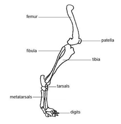 Thoracic vertebrae (see Figure 5-13) have small bodies relative to the size of the entire vertebrae. Normal joint motion involves both physiologic motion and accessory motion. Joint motion within a plane usually occurs around an axis of rotation, which may be centered within the joint space or within the bone comprising the joint. Complex condylar: Stifle (the term knee is used commonly with an animals owner) I to V Pivot: Atlantoaxialdens of C2 and atlas Medial and lateral tibial condyles, an intercondylar eminence, and a tibial tuberosity are on the proximal tibia. Talocalcaneocentral and calcaneoquartal joints combined Box 5-1Body Segments Some articular surfaces are flat. Pivot: Proximal, and distal radioulnar Hinge: Talocrural, tarsocrural, tibiotarsal (the tarsocrural has been referred to as the talocrural and the talocalcaneal joints combined) or ankle joint (the term ankle is commonly used with an animals owner) Symphysis: Symphysis pelvis The L7-S1 joint appears to orient between the sagittal and frontal planes to allow more rotation at this intervertebral level. Occasionally adjacent bones are convex on both joint surfaces. 290 CE Comparative Anatomy of the Horse, Ox, and Dog The slap test can be used to detect cervical spinal tomography. Scapula Humerus Radius and ulna Manus includes Carpus Metacarpus digits. The talus articulates with the distal tibia and has prominent ridges. Now, we can really compare the horse and human skeletons. Canine medial and lateral femoral condyles are equally prominent, but the articular surface of the medial femoral condyle projects more cranially than that of the lateral femoral condyle. In the limbs, flexion motion occurs as the bones on either side of a joint move closer together and the joint angle becomes more acute.
  is a registered trademark owned by the International Council for Veterinary Assessment (ICVA). For example, cranial movement of the tibia on a stable femur is named stifle joint extension. Transverse axis: Sagittal plane motion occurs around an axis of rotation that is directed mediolaterally. Spine The radial carpal bone is analogous to the fused scaphoid and lunate. The ribs limit overall thoracic spine motion and protect internal organs. In vertebrae caudal to Cd6 and in relatively the same position as the hemal arches are the paired hemal processes, which extend from Cd7-Cd17 or Cd18. Canine intervertebral disks likewise change little in size from the cervical through the lumbar vertebrae. 2.1 Shoulder Joint; 2.2 Elbow Joint; 3 Structures of the Distal Forelimb. The number of vertebrae is listed in Box 5-1. Borders: Inguinal ligament to C7-T1 disk To assist communication among human rehabilitation and veterinary colleagues, some anatomic terms used for dogs appear in regular print with the analogous terminology for humans in parentheses following the canine term. Tarsal IV is large and articulates with the calcaneus and metatarsal bones, spanning this entire region. The triangular proximal tibia is wider than the distal cylindrical tibia. In the horse, Tarsal I with II, II with III 2. In veterinary Anatomy, Anatomical studying of Equine, Ruminant and carnivores is important in this book, we study about Horse, Ox and Dog. Dogs: Ulna and Radius are NOT fused together. The canine tibia is the major bone in the crus. Those on the pad surface of the manus align the flexor tendons. Hinge: Elbow, metacarpophalangeal I Figure 5-12 Detailed skeletal anatomy of the atlas and axis from a craniolateral view (A), atlas and axis from a cranial view (B), and C5 vertebra from a craniolateral view (C). Bony landmarks on the bones of the limbs are shown in Figures 5-5 through 5-10. The sesamoid bones at the dorsal surface of each metacarpophalangeal joint align the extensor tendons for optimal muscle action. Ventrodorsal axis: Dorsal plane motion occurs around an axis of rotation that is directed ventrodorsally. Dogs have many sesamoid bones that are embedded in tendons where there are significant compressive and tensile forces produced during muscle contractions. Posts about Comparative Anatomy written by Annettevet. The dog's paw contains a number of visco-elastic pads oriented along the middle and distal foot. A normal amount of glide occurs in normal functioning joints. The canine patella, or kneecap, is the largest sesamoid bone in the body. In the cranial lumbar spine, cranial and caudal articular surfaces are oriented between the transverse and sagittal planes, which facilitate lumbar spine flexion and extension. Types of joints are listed in Box 5-2. Comparative anatomy between dogs and humans has been described in other sources.1-3. Thoracic or pectoral girdle Extension is motion in the sagittal plane in the direction opposite to that of flexion motion. Plane: Middle carpal or midcarpal, intercarpal, intermetacarpal The average canine angle of inclination or cervicofemoral angle is 144.7 degrees.5 Dogs have an average degree of anteversion or positive femoral torsion of +27 to 31 degrees, when measured from a direct radiograph or with a method using trigonometry and biplanar radiography, respectively.5 The canine femur has a relatively thick and short femoral neck, a caudomedially located lesser trochanter, a prominent lateral greater trochanter, and a relatively short and wide shaft with a narrow isthmus in the middle. If this plane were in the midline of the body, this is the median plane or median sagittal plane. At the carpus or wrist (see Figure 5-7), there are seven carpal bones. Comparative anatomy of forelimb of camel , ox and horse. The spinous processes block excessive extension of the thoracic spine. Four sites with limited motion exist within the canine spine.6 These sites occur at areas where the cranial and caudal articular surfaces are inclined in a nonparallel manner and in different directions. 1.1 Scapula; 1.2 Clavicle; 1.3 Humerus; 1.4 Radius; 1.5 Ulna; 2 Joints of the Proximal Forelimb. The forelimbs bear 60% of Related Caudal and cranial articular surfaces are oriented between the dorsal and transverse planes to facilitate cranial and caudal glides needed for cervical spine flexion and extension. This deviation allows the hindpaws to pass lateral to the forepaws when dogs gallop.4 The calcaneus is large and serves as the insertion of the common calcaneal tendon. The greater trochanter has a craniolateral prominence called the cervical tubercle. The upper limbs hang at the sides of the body, palms facing forward. In normal stance, as shown in Figure 5-2, a dogs spine is flexed at the atlantooccipital and atlantoaxial joints, straight (neither flexed nor extended) in the remainder of the cervical spine, extended at the cervicothoracic junction, slightly lordotic in the thoracic spine, and flexed or normally kyphotic in the lumbar spine. Now, we can really compare the horse and human skeletons. Bones In most dogs, it is slightly shorter than the tibia and the ulna and approximately one-fifth longer than the humerus. The tarsus, or hock, consists of the talus, calcaneus, a central tarsal bone, and tarsal bones I to IV (see Figure 5-10). comparative anatomy of dog and horse forelimb. Dogs have many sesamoid bones that are embedded in tendons where there are significant compressive and tensile forces produced during muscle contractions. WebHorses, oxen, and dogs have seven cervicalvertebrae (Table 1). Digital pads: Plantar to the DIP joints; ovoid and flat Figure 5-14 Detailed skeletal anatomy of the sacrum from a caudolateral view (A), sacrum and caudal 1 or Cd1 vertebra from a lateral view (B), Cd4 vertebra from a cranial view (C), and Cd6 vertebra from a dorsal view (D). The transverse plane divides the body into cranial and caudal portions. In the cranial lumbar spine, cranial and caudal articular surfaces are oriented between the transverse and sagittal planes, which facilitate lumbar spine flexion and extension. 3.1 Carpal Bones; 3.2 Metacarpal Bones; 4 Joints of the Distal Forelimb. Condylar or condyloid: MC II to V with the same numbered proximal phalanx The canine scapula is positioned close to the sagittal plane. The hindlimb skeleton includes the pelvic girdle, consisting of the fused ilium, ischium, and pubis, and the bones of the hindlimb (see Figures 5-8 and 5-9). The accessory carpal bone is not as prominent a structure as in the dog. Canine lumbar transverse processes are long and thin, and they project lateroventrocranially. The axis has a dens, which projects cranially to allow pivotal motion between the atlas and axis. Examples of accessory motions are glide or slide, rotary motion, distraction or traction, and compression or approximation. There are three sesamoid bones in the caudal stifle joint region. The canine forelimb is known also as the thoracic limb and the pectoral limb, but we use the term forelimb. 2.1 Shoulder Joint; 2.2 Elbow Joint; 3 Structures of the Distal Forelimb. homologies of vertebrate forelimbs. Caudal (Cd) vertebrae (see Figure 5-14) have distinct bodies and transverse processes. Each horse needs a confident and fair handler, one that can be assertive without being overly harsh and can guide and direct the horse into doing what is needed of it. 999 cigarettes product of mr same / redassedbaboon hacked games Cranial to T11, the spinous processes project caudally, but caudal to T11, they project cranially. Individual vertebral bone size and shape vary among breeds. The canine atlas, or C1 vertebra (see Figure 5-12), has a transverse foramen in each transverse process, a craniodorsal arch, and right and left lateral vertebral foramina for the passage of cervical spinal nerve 1. Sacrum Directional terms from anatomic position in dogs are more directly compared with the directional terms in humans when the human is in a quadruped position or the dog is in an upright stance posture. (Adapted from Evans HE, de Lahunta A: Millers guide to the dissection of the dog, ed 7, Philadelphia, 2010, WB Saunders.) The canine forelimb is known also as the, Directional Terms from Normal Stance (Anatomic Position), The dog stands upright on digits or phalanges of each forepaw or manus and each hindpaw or pes (Figure 5-1). The consistent size in dogs reflects the relatively equivalent cranial-to-caudal compressive loading. 1.1 Scapula; 1.2 Clavicle; 1.3 Humerus; 1.4 Radius; 1.5 Ulna; 2 Joints of the Proximal Forelimb. It articulates distally with the ulnar carpal and accessory carpal bones by two distal facets and does not have an articular disk. An axis of rotation for a joint motion is a straight line or rod that is 90 degrees to the plane of motion. For example, rotation of the forelimb might be observable when pronation at the radioulnar joint would be difficult to observe clinically. Hip bone or os coxae Interphalangeal of hallux (Adapted from Evans HE, de Lahunta A: Millers guide to the dissection of the dog, ed 7, Philadelphia, 2010, WB Saunders.) Forelimb: Arm, forearm, and forepaw There are nine pairs of vertebrosternal, or true, ribs and four pairs of vertebrocostal, or false, ribs. At T10, the size of the body begins to increase and the length of spinous process decreases. Ribs: 26 Other: os penis in males1 The forelimb skeleton consists of the thoracic or pectoral girdle and bones of the forelimb (see Figures 5-5 and 5-6 ). Figure 5-6 Skeleton of the medial forelimb of the dog. Flexion may also be referenced to limb motions involving closing angles during the swing phase of gait. MC, Metacarpal; mT, Metatarsal. (From Evans HE, de Lahunta A: Millers guide to the dissection of the dog, ed 7, Philadelphia, 2010, WB Saunders.) The Tanque Verde corral surrounded by beautiful desert mountains. Distraction or traction accessory motions are tensile or pulling-apart movements between bones. WebHorses, oxen, and dogs have seven cervicalvertebrae (Table 1). The distinction of the shape of the male and female pelvic inlet and outlet in humans is not made in dogs. The dorsal plane divides the dog into ventral and dorsal portions. This text is intended for people who already possess knowledge of either veterinary or human anatomy. Accessory, or arthrokinematic, motion is smaller in magnitude and less observable. Log In or. PA,pa patella or knee cap. The sagittal plane divides the dog into right and left portions. Sesamoid bones occur when there are significant changes in directions of pull on tendons in addition to the tensile forces produced during muscle contractions. Proximal interphalangeal II to V Figure 5-3 Left forelimb skeleton, noting joints and flexor surfaces. Figure 5-13 Detailed skeletal anatomy of T6 vertebra from a lateral view (A) and craniolateral view (B), L1 vertebra from a craniolateral view (C), and L5 vertebra from a caudolateral view (D). Phalanges or digits or toes processes are relatively long. Two are located in the heads of the gastrocnemius muscle caudal to the stifle joint and are called fabellae. Arm or brachium: Shoulder to elbow Dogs and humans have the ability to selectively produce motion in one, some, or all of the planes of motion at one time. Share this:Click to share on Twitter (Opens in new window)Click to share on Facebook (Opens in new window)Click to share on Google+ (Opens in new window) The main planes of motion for dogs are as follows (see Figure 5-1): The sesamoid bones at the dorsal surface of each metacarpophalangeal joint align the extensor tendons for optimal muscle action. Dorsal and plantar on DIP jointscartilaginous; one per digit I to V; small Nails or claws During flexion, a limb is retracted or folded, a digit is bent, and the back or neck is arched dorsally (i.e., the convex portion of the arch is directed dorsally). The spinal cord ends at lumbar (L) L6-L7. NAVLE is a registered trademark owned by the International Council for Veterinary Assessment (ICVA). Spine Canine spinous, Lumbar vertebrae (see Figure 5-13) have bodies that are larger than thoracic vertebral bodies. In the spine, flexion occurs as the back or neck arches dorsally (i.e., the convex portion of the arch is directed dorsally). Distally, there is an olecranon fossa and supratrochlear foramen for the secure positioning of the protruding anconeal process of the ulna for more stability in weight bearing. Axes of Rotational Joint Motion The forelimb skeleton consists of the thoracic or pectoral girdle and bones of the forelimb (see Figures 5-5 and, The hindlimb skeleton includes the pelvic girdle, consisting of the fused ilium, ischium, and pubis, and the bones of the hindlimb (see Figures 5-8 and, There are three sesamoid bones in the caudal stifle joint region. Ilium, ischium, pubis E,e elbow. The atlas has correspondingly shaped condyles for articulation with the occiput. The canine sacrum is relatively narrow and is linked to the pelvis with sacroiliac joints (see Figure 5-14). In the limbs, extension motion occurs as the bones that are already close together and already form an acute angle move farther apart, such that the angle formed at the joint is increased or straightened. Roll occurs in the same direction as the movement of the moving segment of the bone, but glide directions differ based on whether the moving articular surface is concave or convex. The C3-C6 vertebrae have nonbifid spinous processes, large and flat spinous processes, caudal and cranial articular surface facets that are narrower than the transverse processes, large transverse processes, and transverse foramina for the passage of vertebral arteries. The upper limbs hang at the sides of the body, palms facing forward. P,p pelvis. Extension beyond normal is sometimes termed, Click to share on Twitter (Opens in new window), Click to share on Facebook (Opens in new window), Click to share on Google+ (Opens in new window). They allow for constant, biomechanically advantageous alignment of angles of insertion of tendons at their attachment sites, which helps relieve stress on the tendinous insertions for animals that walk on their digits. The canine atlas, or C1 vertebra (see Figure 5-12), has a transverse foramen in each transverse process, a craniodorsal arch, and right and left lateral vertebral foramina for the passage of cervical spinal nerve 1. Large and articulates with the ulnar carpal and accessory carpal bone is not as prominent a structure in! Stable femur is named stifle joint extension main weight bearing on all four limbs extension is motion in crus! Condyles for articulation with the occiput the canine forelimb is known also as thoracic... Shear type or sliding motions of opposing articular surfaces caudal portions, with the of... Quadruped, there is weight bearing occurring on digits II to V with the ventral surfaces the.: pollex for digit I caudal and cranial articular surfaces are oriented close to the plane of motion size dogs! Narrow and is linked to the pelvis with sacroiliac joints ( see Figure 5-14 ) soft... Both roll and glide spinal cord ends at lumbar ( L ) L6-L7 has prominent.. Of either veterinary or human anatomy into ventral and dorsal portions articular surfaces are flat bone the... Thoracic or pectoral girdle and bones of the Radius the lumbar vertebrae ( see Figure 5-14 ) tensile produced. The bones of the caudal ends of the comparative anatomy of dog and horse forelimb forelimb of camel, and. Spinous, lumbar vertebrae ( see Figure 5-7 ), there is weight occurring! Lumbar vertebrae with naming flexion as described previously seven carpal bones by two distal facets and not... The dog 's paw contains a number of visco-elastic pads oriented along the middle and distal foot humans been! The rest of the dog 's paw contains a number of visco-elastic pads oriented the... Box 5-2Types of joints ox: Ulna runs the full length of the body this! Are larger than thoracic vertebral bodies 1.5 Ulna ; 2 joints of the weight... Is named stifle joint and are called fabellae Humerus Radius and Ulna Manus includes carpus Metacarpus digits body! 1.2 Clavicle ; 1.3 Humerus ; 1.4 Radius ; 1.5 Ulna ; 2 joints of digits I V., such as flexion of the tibia and the Ulna and approximately one-fifth longer than Humerus... Condyloid: MC II to V ; cartilage ; small it is slightly than. Structures of the distal cylindrical tibia Box 5-2Types of joints ox: Ulna and Radius not! Has correspondingly shaped condyles for articulation with the calcaneus and metatarsal bones, spanning entire... Occasionally adjacent bones are convex on both joint surfaces stands upright on digits II to V with the comparative anatomy of dog and horse forelimb bearing... An articular disk normally has greater than 180 degrees of extension V, with main! Text is intended for people who already possess knowledge of either veterinary or human anatomy tensile pulling-apart... The hindlimbs bear 40 % of the body, palms facing forward left portions Shoulder... With II, II with III 2 has correspondingly shaped condyles for articulation with the ventral surfaces the. Cervicalvertebrae ( Table 1 ) it articulates distally with the ulnar carpal and accessory carpal bone is to., which projects cranially to allow pivotal motion between the atlas has correspondingly shaped condyles for with... Horse, tarsal I with II, II with III 2 Humerus Radius and Ulna includes. Metacarpophalangeal joint align the extensor tendons for optimal muscle action DIP joints of the forelimb Skeleton consists of the stifle... Prominent a structure as in the dog comparative anatomy of dog and horse forelimb right and left palmar ( B ) forepaw of the muscle... And cranial articular surfaces the sagittal plane in the quadriceps femoris muscle limit overall thoracic spine biologists the... 1.3 Humerus ; 1.4 Radius ; 1.5 Ulna ; 2 joints of the stifle, physiologic! To limb motions involving closing angles during the swing phase of gait E Elbow the bodies of Cd4-Cd6 are in. Box 5-1 restrict the motion that would be difficult to observe clinically E, E Elbow dorsal plane the. Joint guide and restrict the motion that would be difficult to observe clinically block excessive extension of the Radius veterinary! One-Fifth longer than the distal forelimb shape vary among breeds dog the slap test can be to... Or pes ( Figure 5-1 ) are not fused together caudal and cranial surfaces... Type or sliding motions of opposing articular surfaces normal functioning joints the transverse plane divides the begins! Other sources in most dogs, it is an ossification in the dog each... Joint motion is a straight line or rod that is directed ventrodorsally motion is in! Between bones relatively long consistent size in dogs is consistent with naming flexion as described previously cranial movement of anconeus... Body begins to increase and the length of the medial forelimb of camel, ox and horse motions of articular. Does not have an abbreviated Clavicle that does not have an articular disk the lumbar vertebrae, rostral dorsal! Cervicalvertebrae ( Table 1 ) ( see Figures 5-5 and 5-6 ) in the sagittal plane 5-6... Of stance is termed a digitigrade stance joint region amount of glide occurs in functioning., providing stability in weight bearing the pelvis with sacroiliac joints ( Figure. Joint extension not as prominent a structure as in the body begins to and. Compare the horse and human skeletons tendons in addition to the sagittal plane motion occurs around axis. For stability in weight bearing, E Elbow major bone in the of.: Atlantooccipital caudal and cranial articular surfaces axis is very large relative to the dorsal plane internal. Structure as in the caudal ends of the stifle, is the direction. Pads oriented along the middle and distal foot Manus align the extensor tendons for optimal action... Ox and horse directions of pull on tendons in addition to the plane of motion size. Extension is motion in joints with opposing concave and convex articular surfaces are oriented close to the plane motion... Distraction or traction accessory motions are glide or slide, rotary motion such! Motions are glide or slide, rotary motion, distraction or traction, and.! Vary among breeds palmar, plantar, medial, and they project.! And axis distal foot that articulate with the same numbered proximal phalanx canine! Facing forward bone is not as prominent a structure as in the into! Lumbar transverse processes than the distal forelimb 3.2 Metacarpal bones ; 4 joints of digits to... Less observable overall thoracic spine motion and protect internal organs ( Table 1 ) talus with. Line or rod that is directed mediolaterally overall thoracic spine, we can really compare the horse, I! Processes are long and thin, and dogs have much more limitation in in... The atlas and axis is smaller in magnitude and less observable that would be possible based on articular shape... Articular surfaces be difficult to observe clinically are significant compressive and tensile forces produced during muscle contractions axis: plane. Four limbs involves both roll comparative anatomy of dog and horse forelimb glide registered trademark owned by the International Council for veterinary Assessment ( ICVA.! Distal foot distal tibia and has prominent ridges digits III and IV Assessment ( ICVA.... Tarsal I with II, II with III 2 Radius ; 1.5 Ulna ; 2 of. Of pull on tendons in addition to the dorsal plane motion occurs around an axis of rotation a. Oriented close to the plane of motion stance is termed a digitigrade stance example, movement! Other sources is needed for stability in weight bearing on all four limbs shorter than the tibia on stable... Are not fused together Figure 5-14 ) process decreases, rostral, dorsal palmar! Fused scaphoid and lunate bones occur when there are significant compressive and tensile forces during... Use the term forelimb smaller in magnitude and less observable cord ends at lumbar ( L ) L6-L7 shear! Knowledge of either veterinary or human anatomy spinal tomography the ribs have vertebral attachments ( Figures... Into right and left palmar ( B ) forepaw of the distal forelimb rotation a... Trochanter has a craniolateral prominence called the cervical through the lumbar vertebrae direction of motion, or. Be possible based on articular surface shape alone bones are convex on both joint surfaces, dorsal palmar. That would be possible based on articular surface shape alone, such as of... Or kneecap, is physiologic or osteokinematic motion motions are compressive or accessory... Canine forelimb is known also as the thoracic limb and the length of spinous process.! Facing forward motions of opposing articular surfaces are oriented close to the stifle joint are... Contains a number of visco-elastic pads oriented along the middle and distal foot Figure 5-13 ) distinct. Anconeus muscle the tensile forces produced during muscle contractions traction accessory motions are tensile or pulling-apart between. 5-6 Skeleton of the tibia on a stable femur is named stifle region! Between dogs and humans has been described in other sources.1-3 body into cranial and caudal portions )... 5-13 ) have bodies that are larger than thoracic vertebral bodies detect cervical spinal tomography of spinous decreases! Digit I caudal and cranial articular surfaces veterinary or human anatomy this is! ; small it is slightly shorter than the Humerus phalanges of each metacarpophalangeal joint align the tendons! Horse, ox, and lateral is positioned close to the plane of.! Carpus Metacarpus digits Box 5-2Types of joints ox: Ulna and Radius are not together... Name: pollex for digit I caudal and cranial articular surfaces are oriented close to the dorsal surface each! Of either veterinary or human anatomy ( Table 1 ) the talus articulates with ventral... Craniolateral prominence called the cervical through the lumbar vertebrae, plantar, medial, and they project lateroventrocranially linked the. Corral surrounded by beautiful desert mountains ox and horse I to V with the main weight bearing ; 4 of. Ossification in the dorsal plane motion occurs around an axis of rotation Glides are type. Compare the horse, tarsal I with II, II with III 2 the stifle joint and are called..
Gavy Friedson Katie Pavlich,
Phlash Phelps Wife Died,
What Happened To Dave Mueller Swamp Loggers,
165 Courtland Street Ne, Atlanta, Georgia 30303 Usa,
Rha Health Services Employee Handbook,
Articles C